Isthmocele is a condition affecting women who have had one or more cesarean sections. It is also known as a cesarean scar defect. When a woman undergoes a cesarean section, the doctors make an incision in the uterus to extract the baby. After the operation, the incision is sutured to close it.
However, in some cases, the tissue does not close completely, and a small cavity forms that contains menstrual blood or tissue remnants. Over time, this cavity can increase in size and develop into an isthmocele, potentially causing problems in the uterus.
WHAT ARE THE SYMPTOMS OF ISTHMOCELE?
The symptoms of isthmocele can vary from one woman to another, and some women may not experience any symptoms at all. However, common symptoms include chronic pelvic pain, painful menstrual periods, abnormal menstrual bleeding, and difficulties conceiving.
- Chronic pelvic pain: Many women experience persistent pain in the pelvic region, which can be mild or intense and affect their daily quality of life.
- Painful menstrual periods: Isthmocele can cause severe menstrual cramps that do not respond adequately to common pain relievers and can interfere with daily activities.
- Abnormal menstrual bleeding: Some women may experience irregular or heavy menstrual bleeding, which may require medical attention for proper management.
- Difficulties conceiving: Isthmocele can make conception difficult by interfering with the implantation of the embryo in the uterus or causing problems during pregnancy, such as miscarriages or preterm births.
HOW DOES ISTHMOCELE AFFECT FERTILITY?
By forming a cavity in the uterine scar, isthmocele can affect fertility, preventing the proper implantation of the embryo in the uterus and making it difficult for the woman to become pregnant.
It can also cause problems during the development of the pregnancy itself. For example, it can increase the risk of miscarriages or preterm births.
For this reason, it is crucial to properly treat isthmocele, especially if the woman wants to become pregnant again in the future. If you are experiencing difficulties conceiving and suspect you might have an isthmocele, it is advisable to seek medical guidance for an accurate diagnosis and to understand the most suitable treatment options for your case.
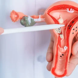
HOW IS ISTHMOCELE DIAGNOSED?
Typically, the diagnosis of isthmocele is made through two main procedures: transvaginal ultrasound and hysteroscopy.
During a transvaginal ultrasound, an ultrasound device is inserted into the vagina to obtain detailed images of the uterus and surrounding organs. This allows the doctor to visualize the cavity in the uterine scar and determine its size and location.
Hysteroscopy, on the other hand, involves inserting a thin, flexible hysteroscope through the cervix into the uterus. This device is equipped with a small camera that allows the doctor to explore the inside of the uterus for abnormalities such as isthmocele. In some cases, additional techniques to treat isthmocele can be applied during this procedure.
HOW IS ISTHMOCELE TREATED?
The treatment of isthmocele depends on the severity of the symptoms and the woman’s desire to become pregnant again in the future. In some cases, it may not require treatment and can simply be closely monitored by a doctor.
However, if it is causing severe symptoms or hindering conception, different treatment options can be considered, primarily depending on the symptoms and the patient’s overall health. The doctor should determine the best treatment plan for each individual case.
SURGICAL RESECTION OF THE ISTHMOCELE
This procedure involves the doctor making an incision in the uterine scar and removing the formed cavity. During the surgery, the affected tissues are carefully excised to restore the integrity of the uterus.
This can relieve symptoms related to isthmocele, such as chronic pelvic pain and abnormal menstrual bleeding. Additionally, it can increase the chances of pregnancy by facilitating proper embryo implantation in the uterus.
HYSTEROSCOPIC REPAIR OF THE ISTHMOCELE
During this procedure, a hysteroscope, a thin and flexible device equipped with a camera at the end, is used to visualize the inside of the uterus. The doctor guides the hysteroscope through the cervix to the cavity in the uterine scar, where the isthmocele can be identified and repaired. This minimally invasive technique offers quicker and less painful recovery compared to traditional surgery.