The uterus constitutes one of the most important parts of the body, as it is an organ belonging to the female reproductive system. The term comes from the Latin “uterus” and is also known as the womb or maternal sac. A woman’s uterus resembles the shape of a pear, is hollow, and is located in the pelvis, between the fallopian tubes and the ovaries. Its size in an adult woman is about 6 to 8 centimeters in length and approximately 5 centimeters in width. In gynecology and assisted reproduction, the uterus is one of the key players that enables embryo implantation and subsequent fetal development.
WHAT IS THE COMMON SHAPE?
The uterus can be divided into the cervix, isthmus, and body. The base is oriented upwards and forwards, while the cervix is slightly directed backwards.
- Cervix or uterine cervix: the lowermost part, located inside the vagina, so named for being elongated and narrow, connecting the vagina and the uterine cavity.
- Body of the uterus: the widest part, located above the cervix. This uterine body consists of two layers. The innermost layer is called the endometrium, which is crucial for embryo implantation. Outside lies the muscular layer or myometrium, with the ability to contract during childbirth.
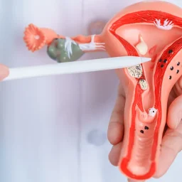
shapes of uterus
CLASSIFICATION OF THE UTERUS ACCORDING TO PHYSIOGNOMY
Congenital anomalies of the uterus are classified depending on the defect produced during the development of this organ in the embryonic period. These specific malformations are also called Müllerian anomalies. A congenital anomaly of the vagina or uterus can reduce a woman’s ability to conceive or prevent the embryo she carries from reaching full term: implantation failures or spontaneous abortions.
Likewise, one of the malignant tumors and enemies of women is the well-known uterine cancer. Although more common in menopausal women, at Ovoclinic, as an assisted reproduction clinic, we advocate prevention to detect any signs of cancer from its onset to avoid major causes. Annual cytology or pap tests, ultrasound, regular gynecological check-ups, and leading as healthy a life as possible are of great help.
According to its physiognomy, the European Society of Assisted Reproduction (ESHRE) classifies uteruses into six categories:
NORMAL UTERUS
It is a hollow cavity, without obstructions, growths, or internal hemorrhages, pear-shaped upside down, with a smaller area at the bottom that connects to the uterine cervix and an upper part surrounded by a smooth muscular wall or zone.
DYSMORPHIC UTERUS
The most common are T-shaped uteruses, whose cavity has this particular shape and not the known triangular shape associated with a normal uterus. It differs from the normal uterus by its walls, which are much thicker, and the cavity is smaller.
shapes of uterus
SEPTATE OR SEPTATED UTERUS
In this type of uterus, there is an internal central wall that divides the uterus into two similar parts. Sometimes, this uterine morphology may be responsible for improper embryo implantation or spontaneous abortions.
BICORNUATE UTERUS
The uterus is “completely” divided into two or well-differentiated parts or zones. Bicornuate uteruses present a total malformation involving the three layers of the uterine wall (endometrium, myometrium, and perimetrium).
UNICORNUATE UTERUS
In this type of uterus, only half of the uterus has developed, so its size is very small compared to a normal uterus.
AGENESIS
shapes of uterus
These are uteruses that have not fully or partially developed. Sometimes, a woman may be born without a uterus.
THE UTERUS DURING PREGNANCY AND POSTPARTUM
During the nine months of pregnancy, the uterus increases in size differentially. A normal uterus (without pregnancy) measures about eight centimeters and weighs about 50-60 grams. When it reaches its maximum size during pregnancy, it can reach two kilograms. Hence, women notice significant changes during the last trimester of gestation.
Similarly, after giving birth, the uterus undertakes the journey in the opposite direction: it shrinks back to its original shape. Afterpains, the contractions that occur after childbirth, help the uterus to regain its size in the days following birth.